Department of Pathology, State University of New York at Stony Brook


Go to:

TOC

Prev

Next

Hodgkins Lymphoma (HL): An Overview

ACTUALLY I fibbed a while back when I said that the great white whale of hematopathology is T-cell lymphomas--it's really Hodgkin lymphoma(Hodgkin's disease and Hodgkin's lymphoma mean the same entity). The disease is named for Thomas Hodgkin, who first described the entity in 1832. For over a hundred years many a medical investigator's career shipwrecked while he or she struggled to harpoon the mystery of this ailment.
        For many decades even its nature as inflammatory, infectious, or neoplastic was unknown. More recently it has been identified clearly as neoplastic, but the cell of origin--lymphoid, histiocytic or other--remained obscure. Only in the past 2 years have molecular probes begun to reveal that the characteristic Hodgkin cell is most likely an aberrant B lymphocyte (for starters the curious can peruse Schwartz RS. Hodgkin's Disease--Time for a Change. N Engl J Med 1997; 337:495-6).
        Besides these puzzles, several features make this neoplasia fascinating:

  • Several morphologically and clinically distinct subtypes exist: diffuse or nodular lymphocyte predominance (5% of cases), nodular sclerosis (60-70%), mixed cellularity (20-30%), and lymphocyte depletion (5%) (1966 Rye Classification).
  • The characteristic cell, the Reed-Sternberg cell, is a bizarre, gigantic cell with more than one large nuclei, each enclosing a large, central, scarlet nucleolus with clear space around it. Finding this cell is a requirement for diagnosis in all sub-types except, perhaps, lymphocyte predominance. Mononuclear variants, sometimes called Hodgkin cells, are also characteristic of the disease but by themselves are insufficiently diagnostic. Other variants of the Reed-Sternberg cell include the lacunar cell (seen in formalin-fixed tissue from nodular sclerosis) and the L&H or "popcorn" cell (seen in nodular lymphocyte predominance).

Reed-Sternberg cells

Lacunar cell

L&H cell

  • A relatively sparse scattering of these bizarre, neoplastic cells is usually accompanied by an overwhelming crowd of benign inflammatory cells including small T lymphocytes, histiocytes, plasma cells, eosinophils, and neutrophils. In fact to make the diagnosis of HL microscopically, two findings are needed: 1) Reed-Sternberg cells and variants and 2) an appropriate inflammatory background. The inflammatory response is ultimately the result of cytokines produced by the tumor cells.
Nodular sclerosis Hodgkin disease 
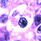
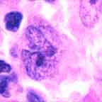
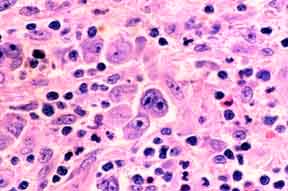
Note the presence both of diagnostic Reed-Sternberg cells and a suitable inflammatory background.

        This entity has also been called Hodgkin lymphoma, but several factors distinguish it from non-Hodgkin lymphoma. This remains true even though its lymphoid origin is increasingly established.


Hodgkin Lymphoma(HL)

Non-Hodgkin Lymphoma (NHL)

Stage / Grade

Because HL begins as a localized process that spreads slowly from one nodal region to another, stage (degree of spread) is important. In some cases grade may be significant, but it is mostly unremarked.

Because NHL is usually systemic from the get-go, grade influences prognosis and therapy more than stage.

Source of Mass

Neoplastic cells are usually <1% of the mass, which is mostly benign inflammatory cells

Almost all the mass is neoplastic lymphoid cells

Immune Deficiency

Usually cell-mediated (T-cell): mycobacterial, fungal, viral, & protozoal infections.

Usually humoral (antibodies from B-cells): bacterial infections.

GI / Waldeyer's Involvement

Rare.

Common.

Marrow Involvement

Significant.

In many cases not important.

Extra-Nodal Involvement

10% of cases

40% of cases.

Treatment

HL is always treated, but milder, localized cases may receive only radiotherapy.

Indolent NHLs may remain untreated for years. Almost all treatment, however, is for systemic disease and thus utilizes chemotherapy. Radiotherapy may be an adjunct.


Epidemiology
In the United States the incidence of HL in recent years has been 2.9 per 100,00, with whites more affected than blacks. In 1998, 7,100 new cases and 1,400 deaths are expected. Mostly because of increasingly effective therapy, the prognosis for HL is good. Currently versus 20-30 years ago, the death rate has declined about 60% and the five-year survival rate is 82% versus 40%.
         The age distribution in developed countries shows 2 modal peaks: young adults (15-34 y) and older adults (50+ y). In developing countries, the young adult peak is replaced by a childhood (0-14 y) form. This difference in pattern between developed and developing countries has suggested to some investigators that exposure to an infectious disease plays a role in HL's etiology. In fact epidemiology, serologic studies, and tissue probes all tend to associate many cases with Epstein-Barr virus. Nonetheless the etiologic role of the virus is controversial, and sensitive techniques are unable to find tissue evidence of the virus in half the cases.

Clinical
Patients usually present with a slowly expanding, non-tender lymph node, mostly cervical, axillary, or inguinal. Only rarely are axial abdominal and pelvic lymph nodes or Waldeyer's ring involved initially. A strange symptom in occasional cases is pain in the involved nodes after alcohol consumption.
        Relapses, especially at untreated sites, tend to maintain the histologic subtype of the original lesion. Generally the natural history of the disease tends toward a progression from forms with few tumor cells and many reactive lymphocytes (diffuse lymphocyte predominance) through an intermediate stage (mixed cellularity) to a terminal state with many bizarre tumor cells and few reactive lymphocytes (lymphocyte depletion). Nodular sclerosis and nodular lymphocyte predominance don't fit as readily into this sequence. The latter has an indolent, relapsing course with a good prognosis.

Extra-Nodal Spread

        In parallel with this progression in subtype is a gradual spread from one lymph node region mostly to contiguous ones. HL originates usually within the neck or mediastinum, only to propagate across the diaphragm along the aortic and iliac lymph nodes and eventually to reach the spleen, liver, and bone marrow. Some cases display "B" or constitutional symptoms such as fever, night sweats and weight loss. Additional morbidity comes from the mass affect of the tumor and impaired cell mediated immunity.
        The outlook would be dismal (a 5 year untreated survival rate of 5%) were it not for effective chemotherapy and radiotherapy that achieve cures in up to 70% of cases. This intensely toxic treatment has its own dangers; second malignancies, most often acute myeloid leukemia, may crop up several years later. To minimize this danger, therapy is tailored to the extent of the disease as determined by a thorough staging. When its results would influence treatment, a staging laparotomy is performed, including splenectomy and biopsies of liver and retroperitoneal lymph nodes. The following staging criteria are used (and in fact may also be used for staging non-Hodgkin lymphoma):

Ann Arbor Staging Classification

Stage I
Involvement of a single lymph node or extra-lymphatic site (IE)
Stage II
Involvement of 2 or more lymph node regions on the same side of the diaphragm or localized involvement of an extra-lymphatic site (IIE)
Stage III
Involvement of lymph node regions on both sides of the diaphragm or localized involvement of an extra-lymphatic organ or site (IIIE) or spleen (IIIS) or both (IIISE)
Stage IV
Diffuse or disseminated involvement of one or more extra-lymphatic organs with or without associated lymph node involvement
The stage can also have a designation of "A" for asymptomatic or "B" for constitutional symptoms.

Table of Contents | Next section | Previous section