|
Nodular Sclerosis Hodgkin Disease
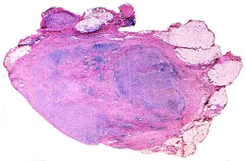
Thick fibrous bands divide this node into cellular nodules containing the neoplastic proliferation. The external fibrous capsule, normally thin and delicate, is also thickened.
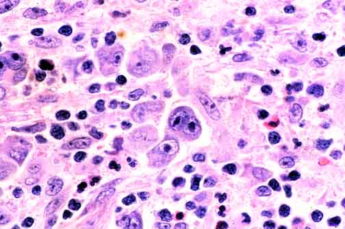
A diagnostic (multi-nucleated) Reed-Sternberg cell lies dead-center. Just below it is a non-diagnostic, uninuclear cell that has been called a "Reed-Sternberg variant" or a "Hodgkin" cell. Although this cell is characteristic of Hodgkin disease, the pathologist who plays by the rules will scrutinize a node suspected of Hodgkin disease involvement until a diagnostic cell is found.
Lacunar cells are a feature of nodular sclerosis Hodgkin disease and are not found in other subtypes. In formalin-fixed tissue, the cytoplasm around Reed-Sternberg cell nuclei retracts, leaving a cleared space possibly spanned by a few shreds of cytoplasm. The nuclei are also contracted and have diminished nucleoli.
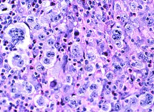
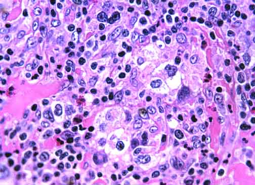
In the midst of this nodule from a different case, sheets of very atypical Reed-Sternberg cells proliferate. This is called the "syncytial variant" of nodular sclerosis Hodgkin disease, and in some studies it has a slightly worse prognosis than cases of nodular sclerosis lacking this feature.

Table of Contents 
|