Go to:

TOC
|
Extra-Nodal Hodgkin Disease
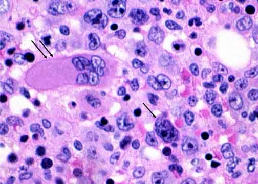
The double arrows point to a megakaryocyte; the single arrow points to a Hodgkin cell. Though clearly different, they can be confused. A diagnosis of marrow involvement by Hodgkin disease can be made if a uninucleated Hodgkin cell is found. Unlike the criteria for a primary tissue diagnosis, finding diagnostic binucleated Reed-Sternberg cells is not required, mainly because the amount of tissue usually available for scrutiny is small. Granulomas and fibrosis are commonly seen in marrows of patients with Hodgkin disease. They are not by themselves diagnostic of marrow involvement, though they should stimulate a thorough search for diagnostic cells.
|
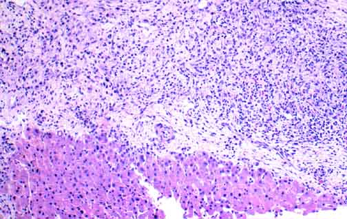
Hepatocytes are at the lower left. Fibrous tissue containing atypical cells is above. At higher power, Hodgkin cells could be identified. The criteria for liver involvement in already diagnosed Hodgkin disease are the same as for marrow involvement.
|

A spleen with 2 nodules of Hodgkin disease. Because splenic involvement may be minimal yet still alter the therapy, it is important to section the spleen as thinly as possible. These sections are usually fixed overnight and then sliced yet more thinly! This way nodules as small as several millimeters in diameter can be found.
|

Table of Contents 
|