 |
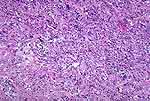
Figure 1: This is a rare malignant fibrous
histiocytoma of the bladder. There is a sprinkling of lymphocytes and some
large, anaplastic nuclei (arrow). Near the center at the far left
(circle), a cluster of cells resembling foamy macrophages, a feature
common to many of these lesions. |
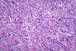 |
Figure 2: Malignant fibrous histiocytoma. Same as
Figure 1. |
 |
Figure 3: Malignant, extrarenal rhabdoid tumor of
bladder. The cells all have eccentric nuclei, and prominent nucleoli, and
some of them have a discrete, pale zone adjacent to the nucleus (arrows).
|
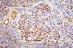 |
Figure 4: Same case as Figure 3, Vimentin
immunostain. The cytoplasm of most of the cells is diffusely positive.
|
 |
Figure 5: Hemangioma of lamina propria. Note that
the blood-filled spaces are not "tightly distended" with blood as would be
seen with passive vascular congestion. |
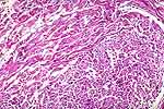 |
Figure 6: Figures 6 and 7 are the high and low
magnifications of neurofibromas in a case of neurofibromatosis. Narrow
strands of collagen with small nuclei (Figure 6) characterize the
neurofibromas. |
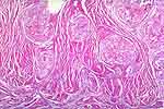 |
Figure 7: This is a low magnification of Figure
6. Note the nodular character of the lesion. |
 |
Figure 8: In a lesion similar to that seen in
Figures 6 and 7, the presence of ganglion cells (circle) would make the
diagnosis ganglioneurofibroma. |