
|
|
| Course
Map: Miscellaneous Tumors: Paraganglioma |
| Paraganglioma | |
| These are similar to those that occur elsewhere; they consist of discrete aggregates of cells ("zellballen") separated by a network of vascular channels. The cells are eosinophilic or dark and, in some cases, their cytoplasm has a lilac color very similar to those of the adrenal medulla. The cells may show some nuclear variation, but this generally will provide no clue as to whether it is benign or malignant. Mitoses are usually absent. These are intramural lesions, with an epicenter usually within the muscularis propria [23]. Where the periphery of the tumor is nearest to the bladder mucosa, one often will see that the cells are smaller and darker, presenting a "neuroblastoma-like" appearance. Most of these patients will have symptoms of catecholamine secretion during micturition (headaches, palpitations, etc). A few of them occur in teens but most are seen in middle life and the sexes are equally affected. About 10-15\% of these are malignant and this determination is made when nodal metastasis or spread to adjacent organs is seen. Bladder paraganglioma is usually a discrete, compact lesion. Widespread dispersion through the bladder wall is reason to suspect malignancy. Pheochromocytoma is a synonym although this term is usually reserved now for those in the adrenal. | |
| Previous Topic | Next Topic |
 |
Figure 1: This is the classic appearance of a bladder paraganglioma. The cells are arranged in aggregates or "zellballen". |
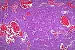 |
Figure 2: This paraganglioma has cells with a tinctorial cytoplasmic appearance very similar to those of the normal adrenal medulla. Note also the prominent vascularity. |
 |
Figure 3: Same case as figure 2. Positive chromogranin. |
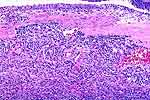 |
Figure 4: This paraganglioma shows in its periphery and near the bladder epithelium at the top, small, dark, "neuroblastoma-like" cells (arrows). These cells are not always present. |
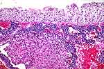 |
Figure 5: This is another paraganglioma, showing an uncommonly prominent layer of "neuroblastoma-like" cells near the bladder epithelium. |
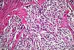 |
Figure 6: This tumor shows the "zellballen" morphology and, in figure 7, it shows widespread dispersion through the muscularis propria. |
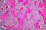 |
Figure 7: Widespread dispersion through the muscularis propria (same case as figure 6). Malignancy should be suspected. This one showed lymph node metastasis. |