 |
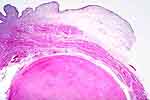
Figure 1: Leiomyoma. Smoothly rounded contours
such as we see here, are not usually present in leiomyosarcomas. |
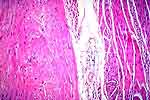 |
Figure 2: Same case as Figure 1. Notice that the
character of the cells in the leiomyoma is not significantly different
from that of the normal muscularis propria on the right side of this
field. |
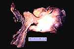 |
Figure 3: Leiomyosarcomas of the bladder often
present as polypoid masses projecting into the bladder lumen. However,
this may be seen also with other spindle cell lesions such as spindle cell
carcinoma and myofibroblastic tumors. |
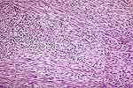 |
Figure 4: Unlike most sarcomas, leiomyosarcomas
usually show long, sweeping, discrete fascicles (arrows) of spindle cells
(except for the very high grade tumors). |
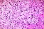 |
Figure 5: Leiomyosarcoma. The fascicular
morphology is more subtle in this case than that in Figure 4, but nuclear
pleomorphism requires a diagnosis of sarcoma. |
 |
Figure 6: This is a transurethral resection of a
bladder tumor. On the left, it would be a problem distinguishing between
leiomyoma and leiomyosarcoma . On the right, it is clearly anaplastic. We
show this photograph in order to stress that a sampling of a part of a
smooth muscle tumor may show a misleading histiology.
|