 |
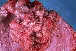
Figure 1: The polypoid gross appearance is
typical of sarcoma botryoides. |
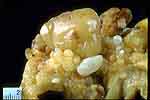 |
Figure 2: Rhabdomyosarcoma, bladder neck. |
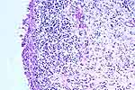 |
Figure 3: The polyps illustrated in Figures 1 and
2 will usually show a concentration of small, dark - staining,
undifferentiated cells forming a band immediately beneath the surface
urothelium (the cambium layer). The deeper tissue is edematous, with
widely scattered tumor cells. |
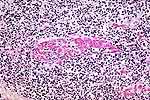 |
Figure 4: A fascicle of the muscularis propria is
seen in the center of the field. It is invaded by small dark tumor cells.
In a pediatric patient or young adult with a bladder neck tumor,
rhabdomyosarcoma would be suspected. On the far left, a cell has a wisp of
pink cytoplasm (rhabdomyoblast--arrow). The diagnosis is rhabdomyosarcoma.
|
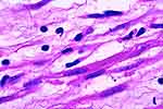 |
Figure 5: This shows a strap cell near the center
of the field. It shows cross striations (arrow). In most cases, these are
not seen, especially in biopsies, and the diagnosis will require
immunohistiochemistry. |