| Injury |


|
| Injury |
|
|
|
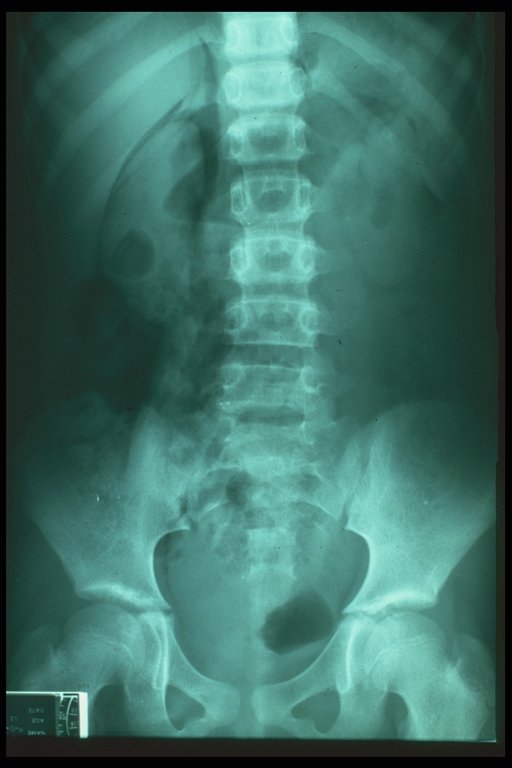
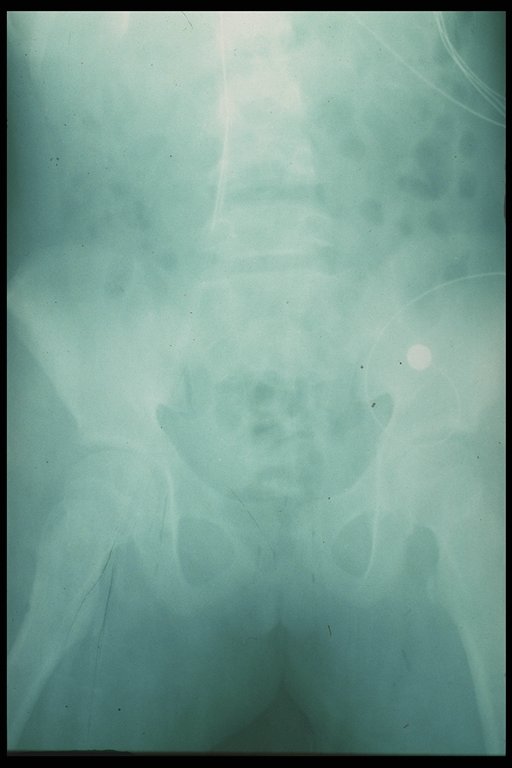
Plain abdominal X-ray in ruptured duodenum, the free air in the retroperitoneum outlined the kidney is shown. |
|
Plain abdomial X-ray in ruptured duodenum, the free air in the retroperitoneum outlined the kidney is shown. |
|
|
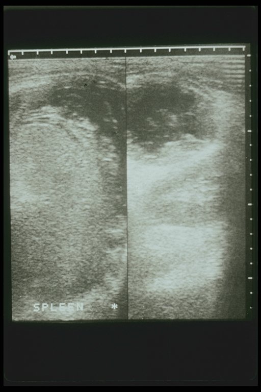
Splenic hematoma is demonstrated by ultrasonography |
|
|
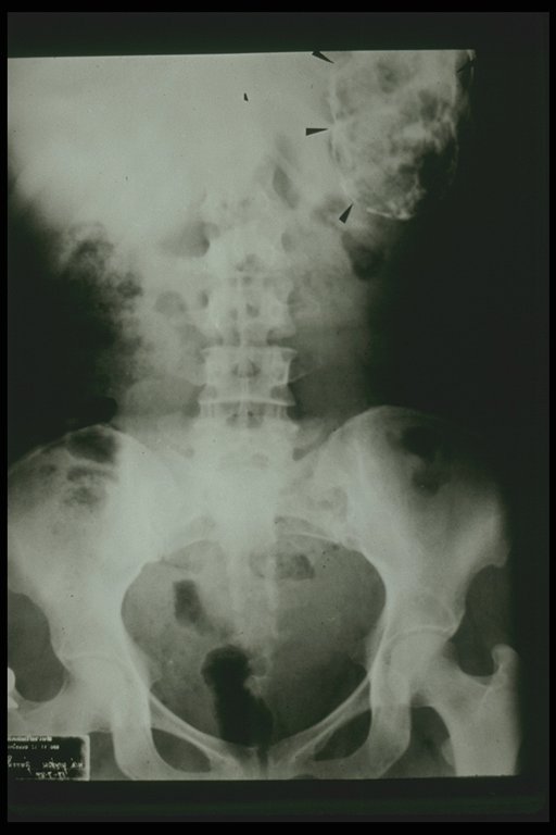
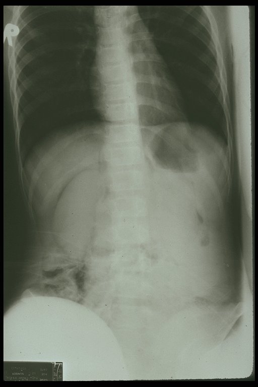
Calcified splenic hematoma. Plain X-ray showed calcification at left upper quadrant of abdomen (arrow) |
|
|
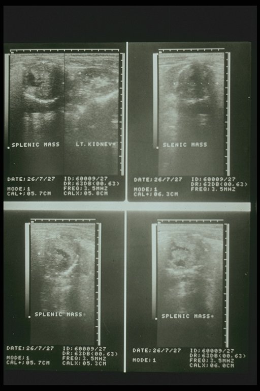
Intrasplenic hematoma demonstrated by ultrasonography |
|
|
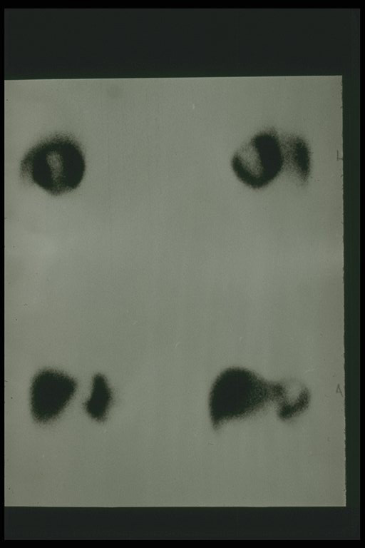
Splenic scan showed low uptake indicating subcapsular hematoma of spleen |
|
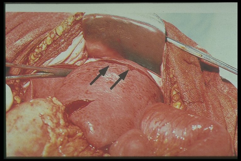
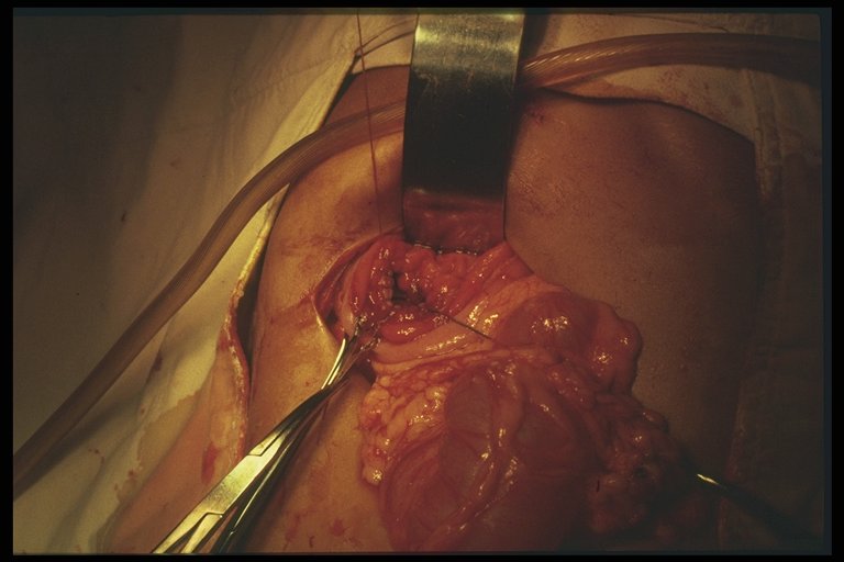
Operataive finding of multiple linear laceration of spleen |
|
|
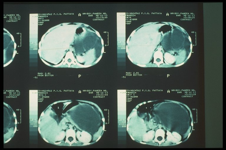
Post-traumatic pancreatic peudocyst demonstrated by CT scan |
|
Pancratic Cystogastrostomy is a procedure of choice when the conservative treatment had failed |
|
Retained guidewire in the Inferior vena cava after angiography (film abdomen) |