| Injury |


|
| Injury |
|
|
|
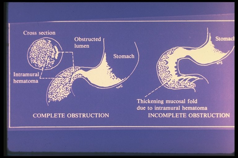
Diagram showed the intramural hematoma of the duodenum (IHD) caused obstruction and explained the finding in contrast study |
|
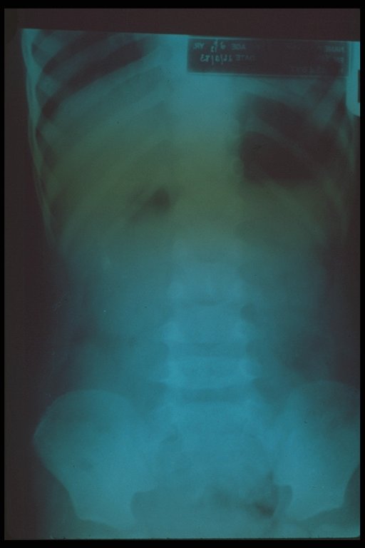
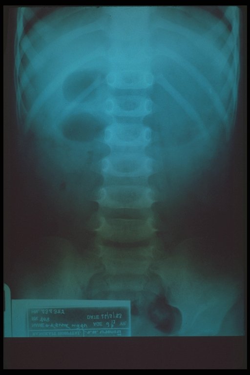
Plain supine abdomen X-ray in case of intramural hematoma of the duodenum showed dilatation of stomach and duodenum |
|
|
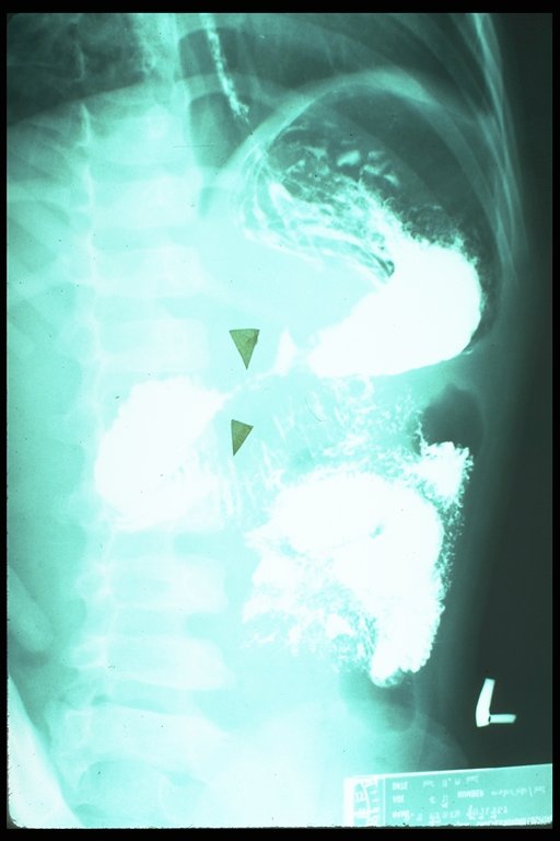
Plain upright abdominal X-ray in case of IHD showed air fluid level in the dilated stomach |
|
|
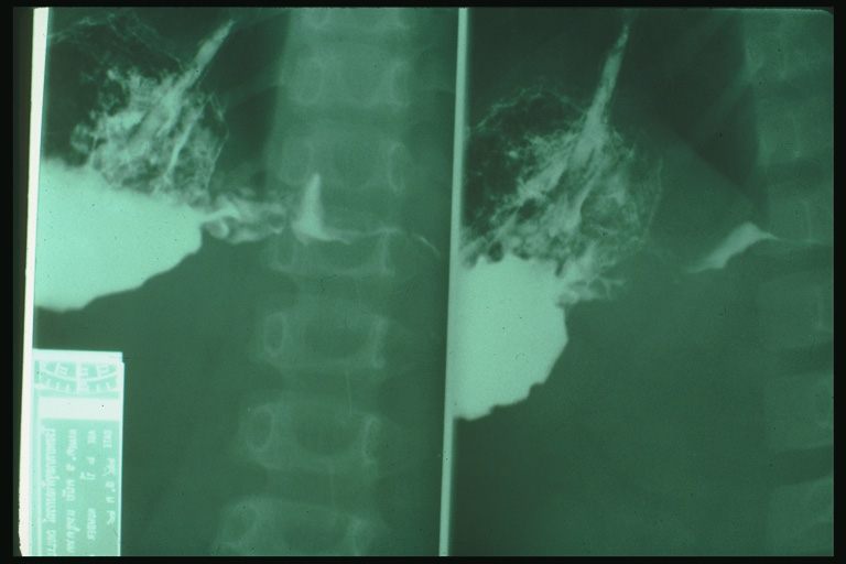
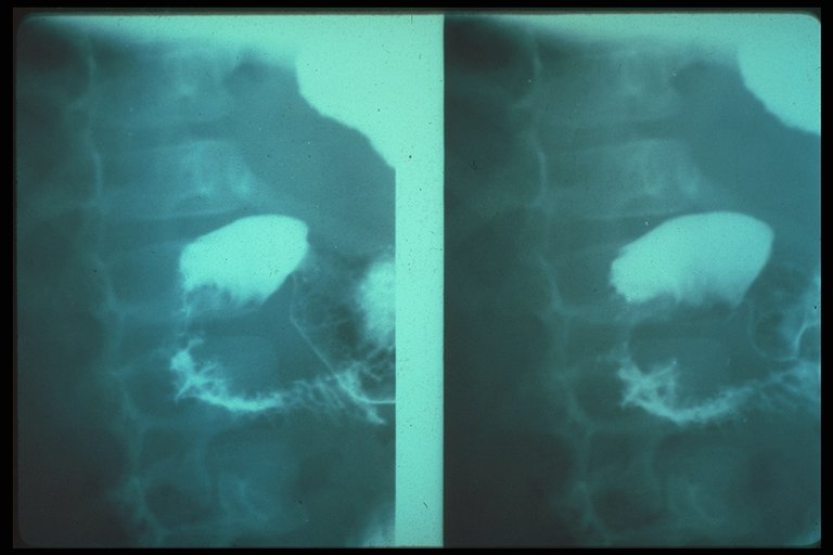
Upper gastrointestinal (UGI) study showed complete obstruction at the duodenim with typical tapering due to intramural hematoma |
|
|
UGI study demonstrated the obstruction at 2nd to 3rd part of duodenum with typical tapering due to intramural hematoma |
|
|
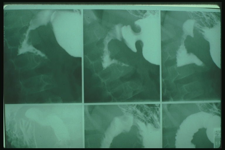
UGI study demonstrated the obstruction at 2nd to 3rd part of duodenum with typical tapering due to intramural hematoma. Stacked coins appearance on the duodenal wall is also noted. |
|
UGI study in IHD showed stacked coins appearance which is a typical finding |
|
|
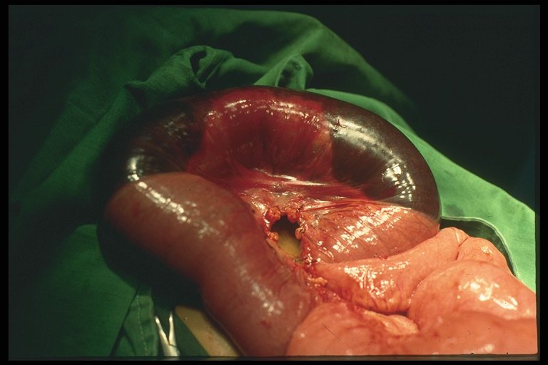
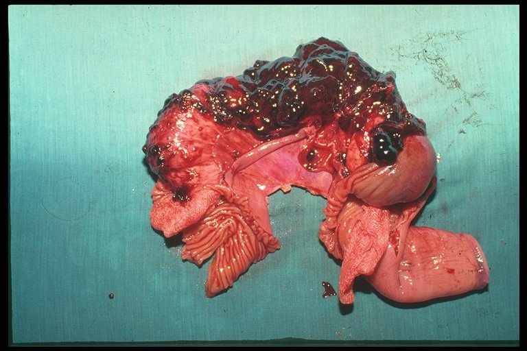
Operative finding of intramural hematoma of the intestine, external appearance |
|
In a resected specimen of massive intramural hematoma, blood clot occurred in the intramural part of intestine |
|
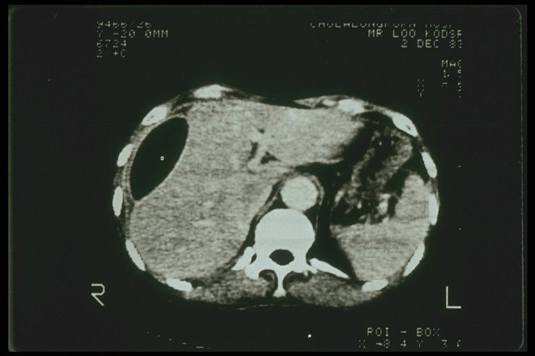
Subcapsular hematoma of the right lobe liver demonstrated by CT scan |