| Abdominal Pain |


|
| Abdominal Pain |
|
|

|
Twisted duplication cyst at transverse colon |

|
Rectal duplication. A barium enema demonstrates hollow mass anterior and connected to the rectum (barium outlined the mass) |
|
|
Rectal duplication. Operative finding of bulging cystic mass into the rectum |
|
|
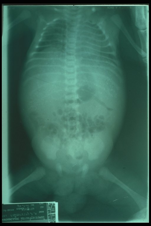
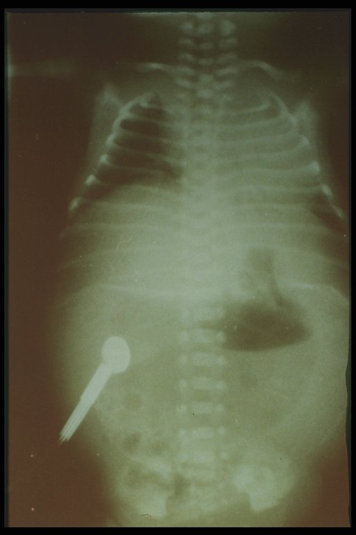
Meconium peritonitis. A classical plain abdominal X-ray demonstrates calcified extravasated meconium in the peritoneal cavity since fetal growth. Free air in the abdomen (football sign) is also noted |
|
|
Meconium peritonitis. A classical plain abdominal X-ray demonstrates calcified extravasated meconium in the peritoneal cavity since fetal growth. Free air in the abdomen (football sign) is also noted |
|
|
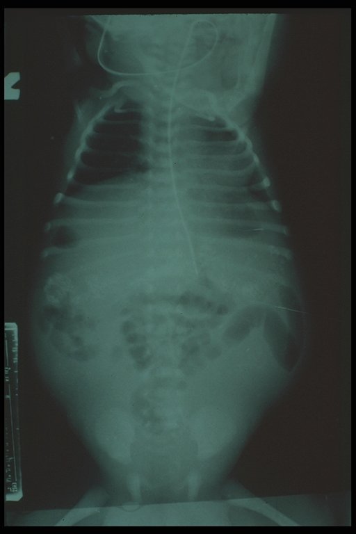
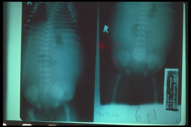
Meconium peritonitis. Scattered concentric pattern of calcification spread throughout the peritoneal cavity with intraabdominal fluid shows that there was meconium leakage sometime during fetal life. |
|
Meconium peritonitis. Scattered concentric pattern of calcification spread throughout the peritoneal cavity with intraabdominal fluid shows that there was meconium leakage sometime during fetal life. |
|
|
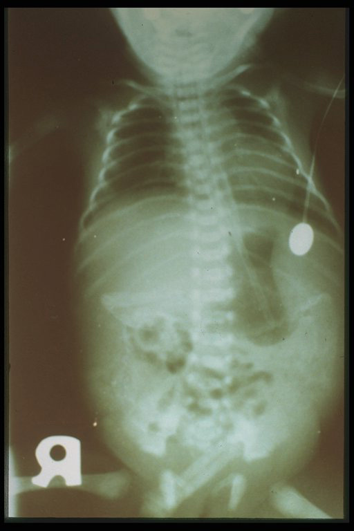
Meconium peritonitis. A classical abdominal X-ray demonstrates calcified extravasated meconium in the peritoneal cavity since fetal growth. Free air in the abdomen (football sign) is also noted. |
|
Meconium peritonitis. A classical abdominal X-ray demonstrates calcified extravasated meconium in the peritoneal cavity since fetal growth. Free air in the abdomen (football sign) is also noted. |
|
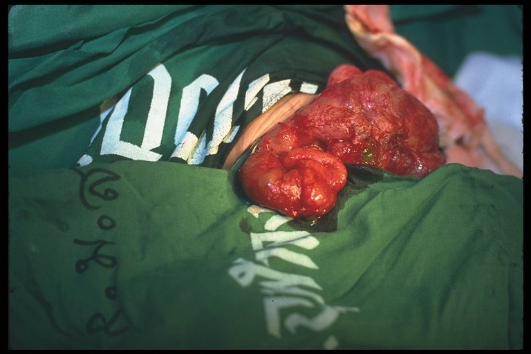
Meconium peritonitis. At operation, matted loops of intestine with calcification over the surface are shown. The perforated site of intestine is also demonstrated where there is a leak of meconium. |