| Respiratory Problem |


|
| Respiratory Problem |
|
|

|
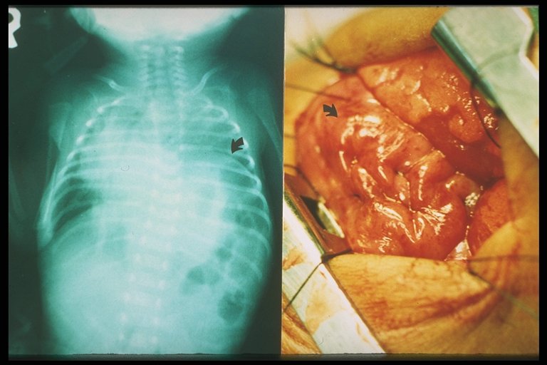
After completion of the plication of diaphragm, the diaphragm is strengthened and flattened |
|
After completion of the plication of diaphragm, the diaphragm is strengthened and flattened flattened (right) and chest X-ray showed that the affected diaphragm was at the normal level. |
|
|
After completion of the plication of diaphragm, the diaphragm is strengthened and flattened flattened (right) and chest X-ray showed that the affected diaphragm was at the normal level.After completion of the plication of diaphragm, the diaphragm is strengthened and flattened flattened (right) and chest X-ray showed that the affected diaphragm was at the normal level. |
|
|
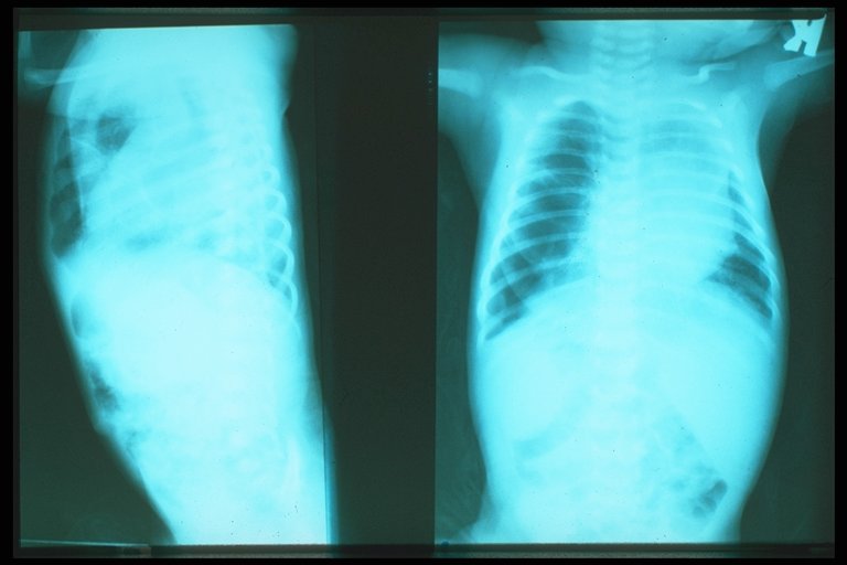
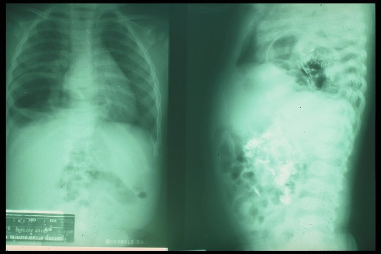
Plain chest X-ray AP (left picture) and lateral ( right picture) showcd soft tissue mass on the right chest cavity with well defined border (soft tissue mass shadow) in a child with hiatal hernia |
|
|
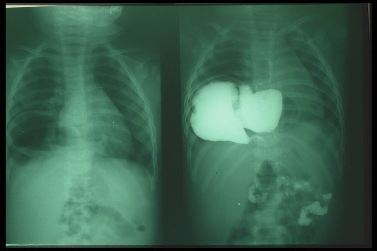
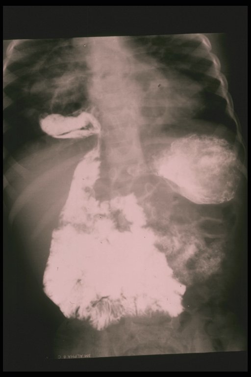
Contrast study outlined the stomach which is herniated into the chest cavity (soft tissue mass shadow ) in a child with hiatal hernia |
|
|
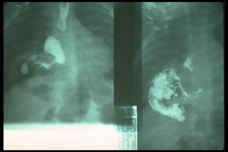
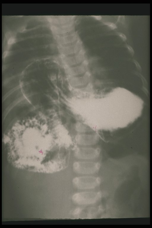
Plain X-ray (right ) and contrast study (left) in a child with right-sided diaphragmatic hernia with sac, the contrast outlined the bowel in the right thoracic cavity |
|
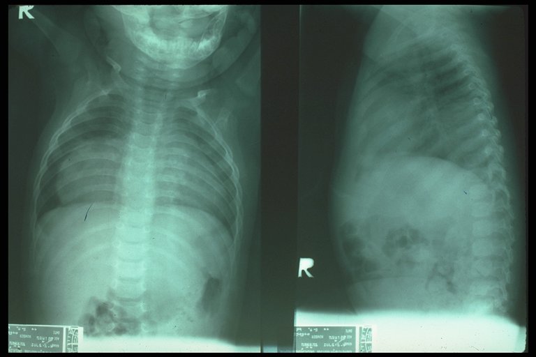
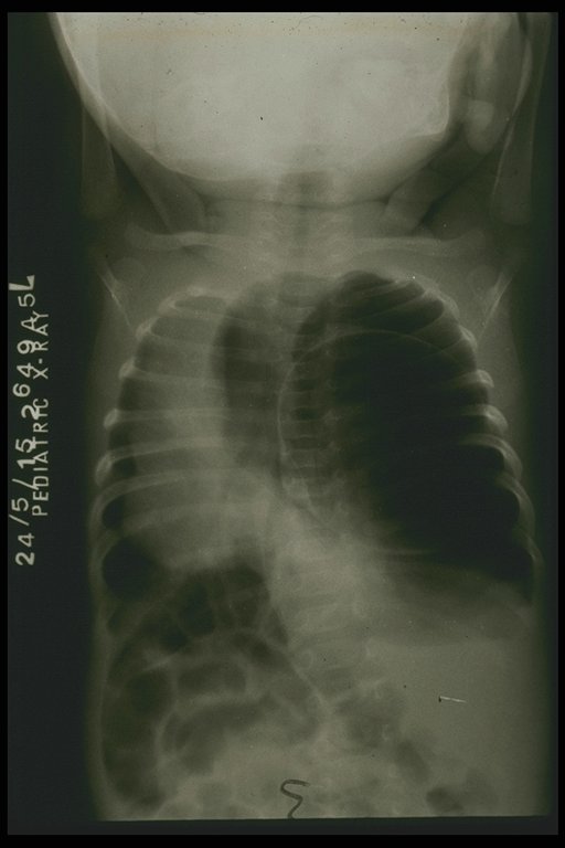
Chest PA film showed abnormal gas over the dome of right diaphragm, chest lateral film confirmed that the abnormal gas was at posterior space. Right posterolateral diaphragmatic hernia was the final diagnosis in this case. |
|
|
Contrast study confirmed the protusion of bowels into the chest cavity in right Bochdalek hernia |
|
Contrast study showed that the duodenum was herniated into the chest cavity, malrotation was also noted. |
|
Abnormal radiolucent area over the left lung field with mediastinal shift to the right in a case of lung cyst with pneumothorax |