| Respiratory Problem |


|
| Respiratory Problem |
|
|
|
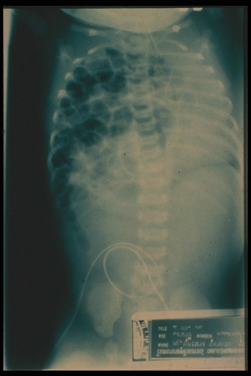
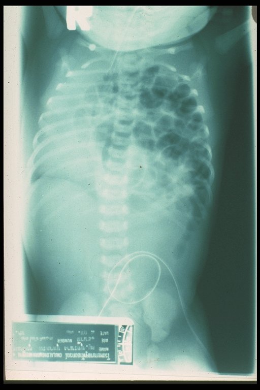
Typical radiographic finding in diaphragmatic hernia. The bowel gas pattern is noted in the left (common side) thoracic cavity and the mediastimum is shifted to the opposite site. |
|
Typical radiographic finding in diaphragmatic hernia. The bowel gas pattern is noted in the left (common side) thoracic cavity and the mediastimum is shifted to the opposite site. |
|
|
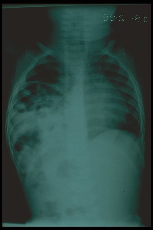
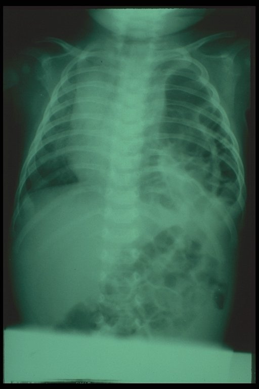
X-ray finding in the right diaphragmatic hernia, uncommon affected side |
|
|
X-ray finding in the right diaphragmatic hernia, uncommon affected side |
|
|
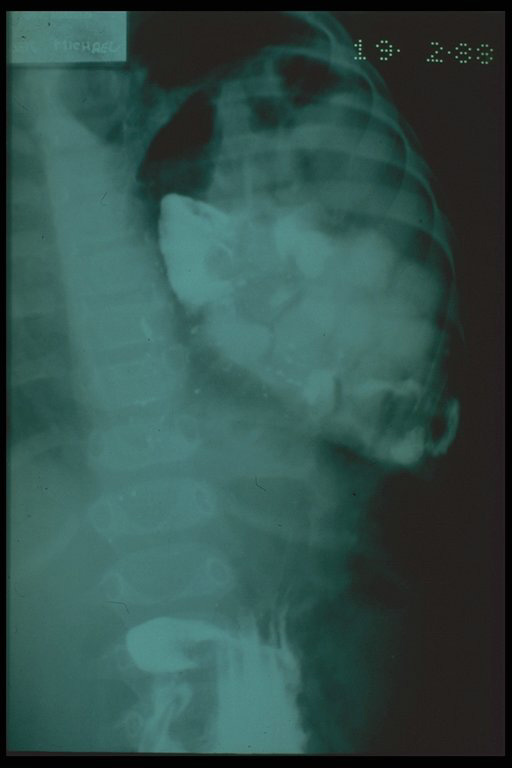
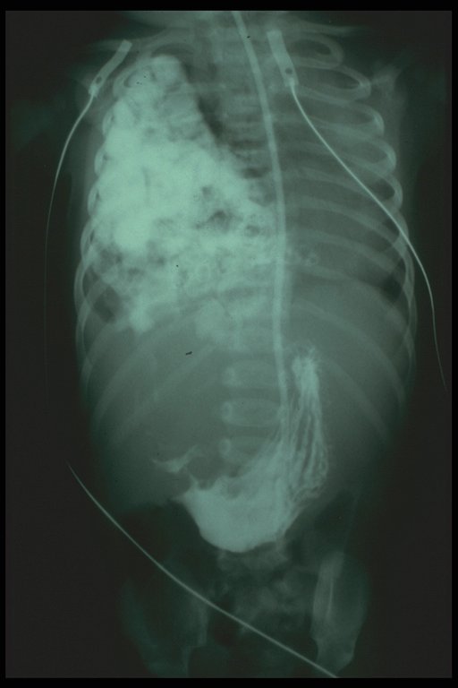
Contrast study was done and the bowels in the thoracic cavity was confirmed |
|
|
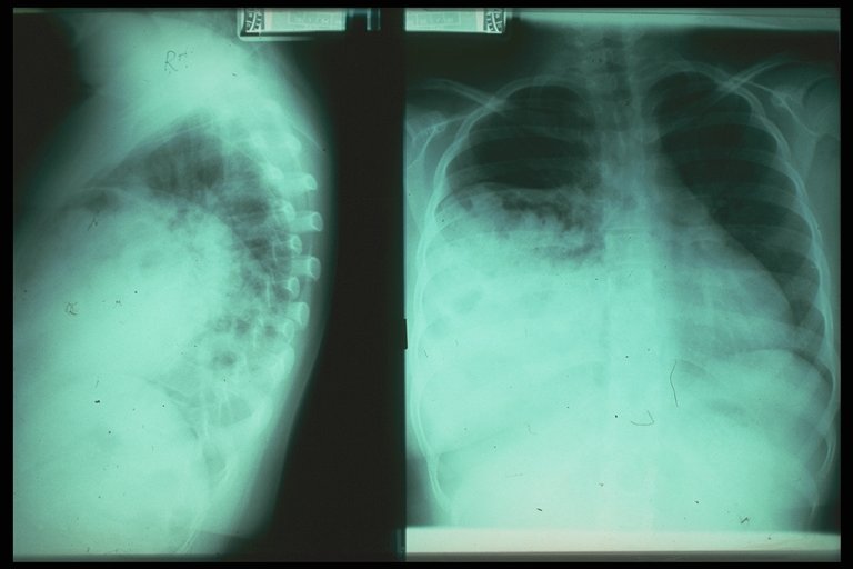
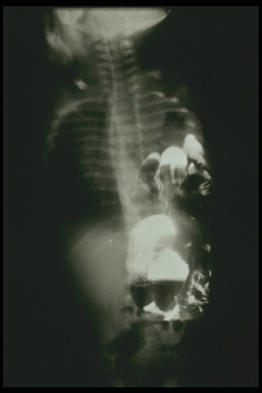
Unusual picture of radiographic finding in diaphragmatic hernia on the right side. The lung shadow in still noted on the affected side and the diagnosis con not be easily convinced by plain chest X-ray . Contrast study is recommended. |
|
Contrast swallowing confirms the diagnosis of right diaphragmatic hernia |
|
|
Contrast swallowing confirms the diagnosis of right diaphragmatic hernia |
|
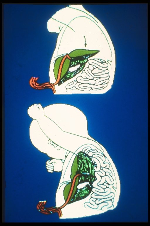
Diagram showed the surgical management. The herniated bowel is reduced from the thoracic cavity and the defect is repaired. |
|
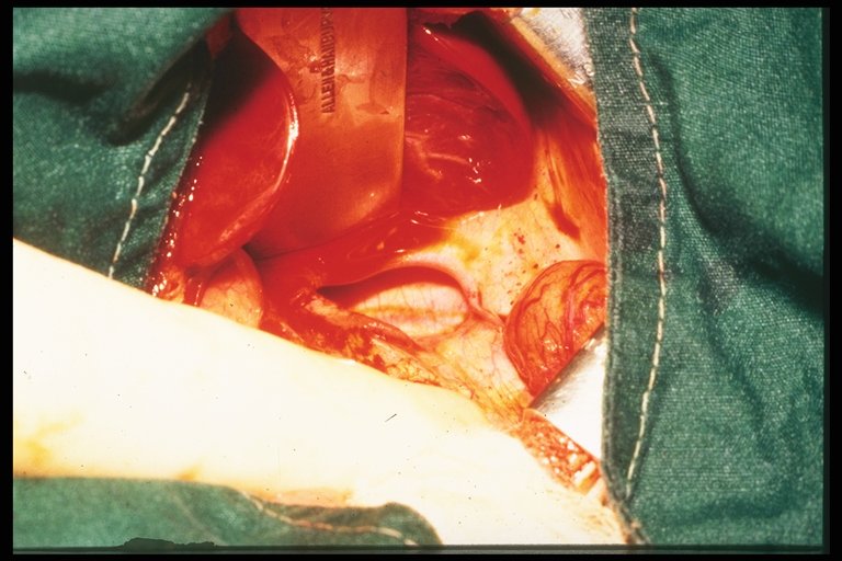
Operative finding in left bochdalek diaphragmatic hernia. A defect at posterolateral part of diaphragm is demonstrated. |