| Abdominal Mass |


 |
| Abdominal Mass |
|
|
|
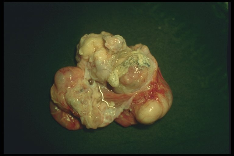
Gross pathology of mesenteric teratoma, mixed tissue with some well-formed calcification are demonstrated |
|
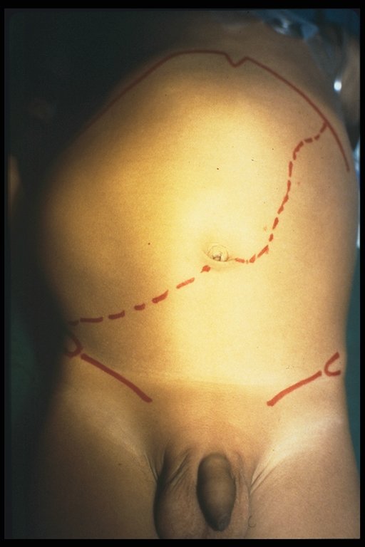
Clinical presentation of hepatoblastoma - right upper quadrant mass |
|
|
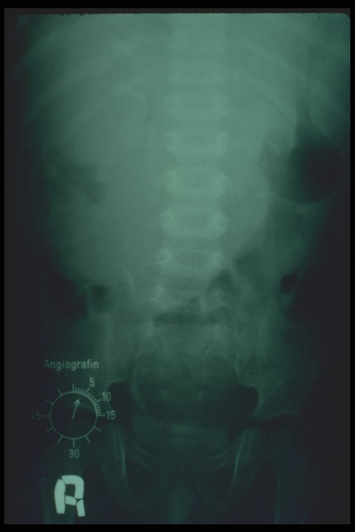
Only right upper quadrant mass is demontrated without specific characters on plain abdominal X-ray in case of hepatoblastoma |
|
|
Ultrasonography of hepatoblastoma |
|
|
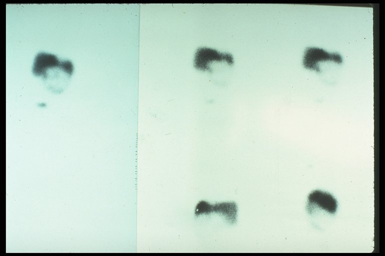
Liver scan in case of hepatoblastoma involved medial and lateral segment of left lobe |
|
|
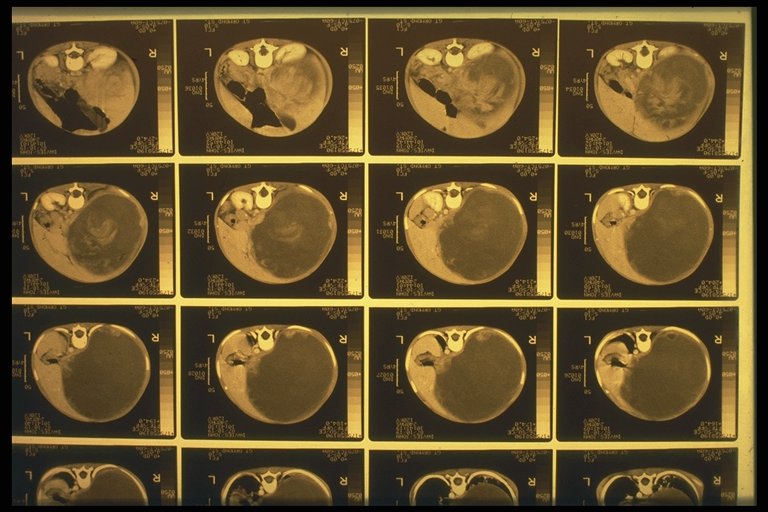
CT scan of hepatoblastoma involved the whole left lobe |
|
|
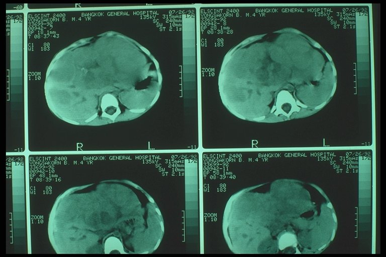
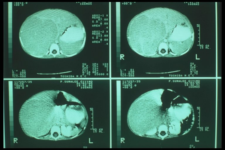
CT scan of hepatoblastoma involved the right lobe |
|
|
CT scan of hepatoblastoma involved the right lobe |
|
|
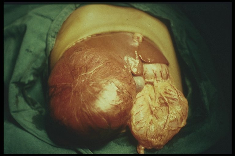
Operative finding of hepatoblastoma involved the right lobe |
|
|
Operative finding of hepatoblastoma involved the dome of right lobe liver |