| Abdominal Mass |


 |
| Abdominal Mass |
|
|
|
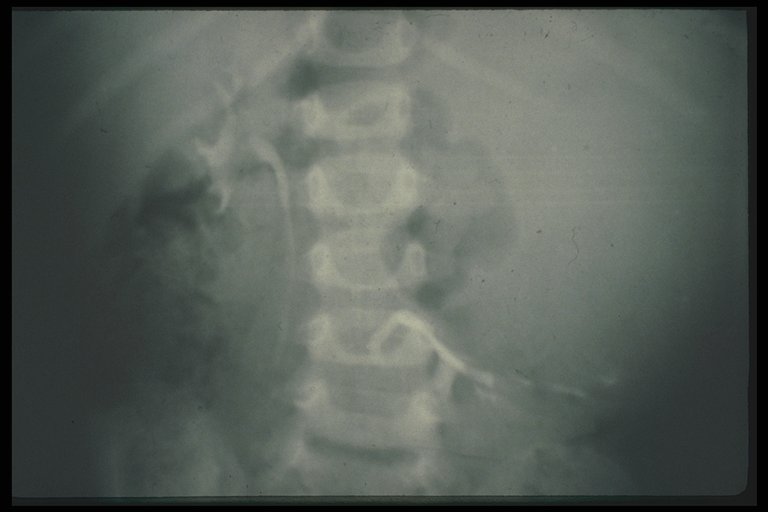
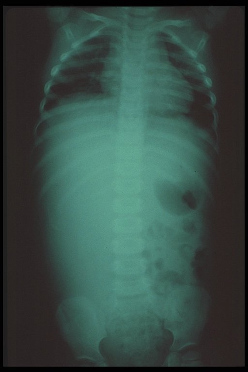
Plain X-ray revealed rim calcification of the Wilms' tumor (arrow) |
|
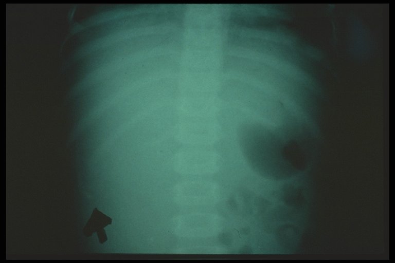
Close-up picture of a plain X-ray of Wilms' tumor showed typical rim calcification (arrow) |
|
|
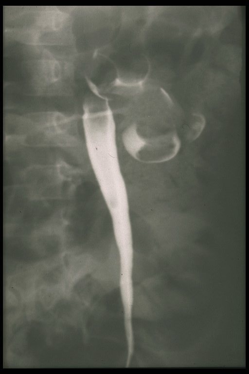
Intravenous pyelography (IVP) showed distorsion of pelvicalyceal system in left kidney - Wilms' tumor |
|
|
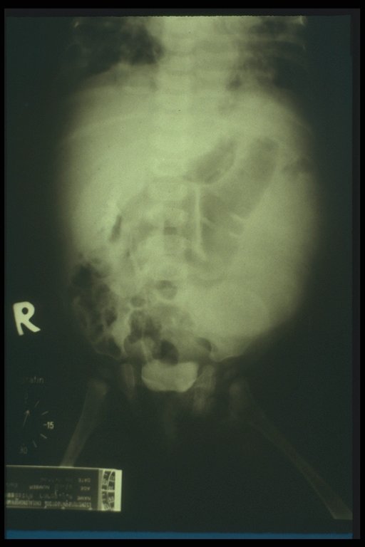
Intravenous pyelography (IVP) showed masses in the pelvicalyceal system, Wilms' tumor is the most likely diagnosis |
|
|
Wilms' tumor, IVP showed non functioned left kidney |
|
|
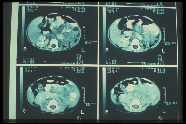
Wilms' tumor, CT scan, showed a huge tumor mass with necrotic tissue in the mass. |
|
Wilms' tumor, CT scan |
|
|
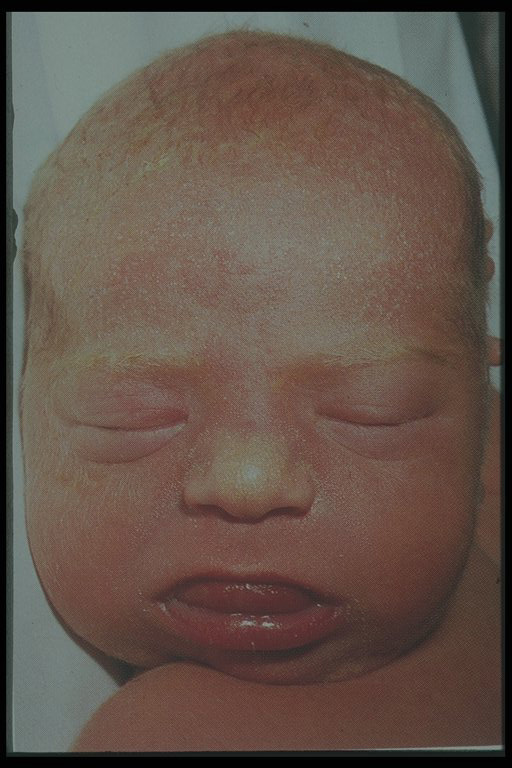
Wilms' tumor associated with Beckwith-Wedemann syndrome |
|
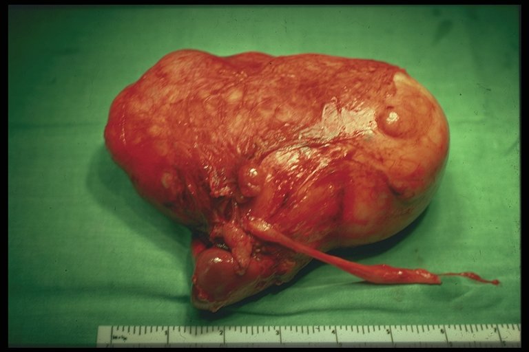
Gross pathology of Wilm's tumor, nephrectomized specimen |

|
Cut surface of specimen, Wilms' tumor at the upper pole displaced the rest of normal kidney tissue downward and outward |