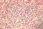
| Aggregates of eosinophilic macrophages (von Hansemann histiocytes)
form nodules or plaques, usually multiple, in the lamina propria [34,67].
There are lymphocytes and plasma cells also present (Fig. 1), but most of
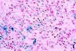
the cells are large and pink-staining. A variable number of them contain
within the cytoplasm the diagnostic Michaelis-Gutmann inclusions (Fig. 2).
These are round 5-8 micron bodies, blue or gray in color. More fully developed
inclusions may assume a targetoid or bull's-eye appearance (Fig. 3) which
may be highlighted with iron or calcium stains (Fig. 4). |