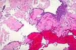
| This is usually a solitary lesion within the lamina propria
and, occasionally, also the superficial zone of the muscularis propria [52,101].
It is an ulcerated and nodular or polypoid lesion which bleeds readily and,
cystoscopically, is not distinguishable from carcinoma. On the microscopic
slide the amyloid material presents a fragmented or "shattered" appearance
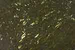
(Fig. 1, Fig. 2, Fig. 3). With polarization of the Congo-red sections there
is an apple-green birefringence (Fig. 4). Bladder amyloidosis is almost
invariably a localized process - unassociated with systemic disease. |