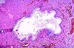
| This consists of endometrial glands and stroma in the bladder
wall identical to endometriosis as seen elsewhere (Fig. 1). Thickening of
the bladder wall (usually the posterior wall), a palpable supra-pubic mass
or cystic, hemorrhagic or edematous mucosal changes can cause confusion
with bladder neoplasia. Microscopically, some of the glands may lack a stromal
element, and some foci may show evidence of recent or remote hemorrhage
[5,28,31,94].
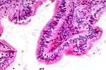
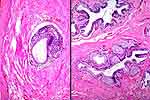
The lamina propria, muscularis propria, or serosa may be involved. A similar
distribution of glands may show the features of the endocervix (endocervicosis;
Fig. 2, Fig. 3, Fig. 5). These glands occur as rounded or stellate-shaped
structures scattered randomly in the bladder wall. The epithelium consists
of a row of cuboidal or columnar cells with pale, mucin-positive cytoplasm.
Least commonly, glandular inclusions may assume a tubal-type morphology
with cilia (endosalpingiosis; Fig. 4). |