
|
|
| Course
Map: Epithelial Tumors: Malignant: Non-urothelial
Carcinoma: Urachal Carcinoma |
| Urachal Carcinoma | |
| The vast majority of these are adenocarcinomas, although they rarely have been urothelial or squamous [93]. The tumors do not differ histologically from those described under primary bladder adenocarcinoma, but most of them are the mucinous (colloid) type. There are no special stains or immunostains that will distinguish urachal from bladder or colonic carcinomas. The urachal carcinoma arises from urachal remnants so they will be located in the bladder dome or adjacent anterior bladder wall, with an epicenter in the muscularis propria rather than the lamina propria. Some may arise from remnants above the bladder and may even be present in the umbilical area. If the biopsy shows benign glandular or intestinal metaplasia in or near the bladder surface, this would point to a primary bladder adenocarcinoma. If similar benign glandular tissue is present deeper in the bladder wall, it likely represents benign urachal remnants, favoring a diagnosis of urachal carcinoma. Again, one must always consider the possibility of a metastasis from the bowel. We have seen several examples of a colonic carcinoma which invaded down through the dome of the bladder. | |
| Previous Topic | Next Topic |
 |
Figure 1: Most urachal carcinomas will have this appearance: fragments of adenocarcinoma (Fig. 1) or signet ring cells (Fig. 2), floating in large lakes of mucin. |
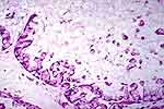 |
Figure 2: Urachal carcinoma. Signet ring cells floating in large lakes of mucin. |
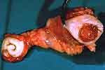 |
Figure 3: This illustrates a typical surgical specimen from a case of urachal carcinoma. The tumor (right) protrudes into the bladder lumen at the dome. The entire urachal tract has been excised, up to and including the umbilicus (left side). |
 |
Figure 4: This specimen shows bladder and bladder neck at the right and a large, necrotic, and mucoid tumor at the left, above the bladder. |
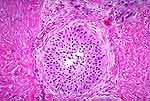 |
Figure 5: This is a urachal remnant, composed of urothelium, deep within the muscularis propria. |
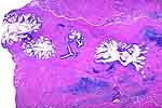 |
Figure 6: Muscularis propria is seen at the top and bottom. Running through it, in a linear fashion, are benign urachal remnants. Most show glandular metaplasia but several (arrows) are lined by urothelium. Obviously, had a tumor been present, we would suspect a urachal origin. |