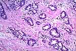
| To make this diagnosis, all of the tumor should be an adenocarcinoma,
with no urothelial or squamous elements [53,100].
Most of them have a resemblance to large bowel carcinomas and, in many cases,
it will be necessary to specifically rule out a colonic or rectal primary.
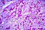
They may be glandular (well-differentiated) or of the signet ring cell type
(poorly differentiated) [18,21,48].
Others may consist largely of mucinous lakes (the mucinous or colloid type)
[75].
They arise from glandular metaplasia of urothelium. If areas of cystitis
glandularis or intestinal metaplasia are present (see below), this would
point to a primary bladder carcinoma rather than a colonic metastasis. This
is true, also, where the bulk of the tumor mass appears to be concentrated
near the luminal aspect of the bladder wall-rather than in the serosal or
deep muscularis propria region. |