| Most of these are seen in the female urethra, but a few occur
in the bladder and in males [29,41,115].
In the older literature they were often classified as mesonephric carcinomas,
although it is not established if any of them arise from mesonephric remnants
[39].
We know that a few of them have arisen from bladder endometriosis, endocervicosis
and endosalpingiosis indicating a mllerian origin [29].
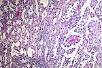
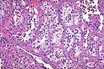
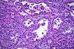
In any case Clear Cell Adenocarcinoma is the term which is now widely used,
even though their designation as "clear cell" leaves something to be desired.
In many of the tumors the cells are brightly eosinophilic as will be seen
in the photomicrographs below. The morphology of the tumor is basically
tubular rather than glandular and there often are papillary and microcystic
areas. The individual epithelial cells often project into the lumen with
a hobnail appearance. The cells have glycogen and, often, mucin in the cytoplasm
or lumina. In the differential diagnosis, we have already noted that the
clear cell variant of urothelial carcinoma does not form tubules. Metastatic
renal cell carcinoma will not have cytoplasmic mucin. Metastatic clear cell
carcinoma of the female genital tract must always be specifically ruled
out. The nephrogenic adenoma causes the most problems in differential diagnosis.
These often have basement membranes around the tubules, they consist of
a single layer of bland cells without mitoses and without cellular stratification.
The NA is benign, and will be discussed later. |