Help on Using This Course Select Course Provide Feedback Online Urologic Pathology: Bladder Course Brochure Request CME for this course The AFIP Homepage
Course Map: Epithelial Tumors: Malignant: Non-urothelial Carcinoma: Small Cell Carcinoma


Small Cell Carcinoma

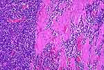
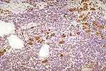
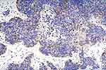
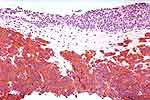
In most cases, these small cell neuroendocrine carcinomas are associated with transitional cell carcinoma in-situ, and, sometimes, with areas of squamous or glandular metaplasia so we would have been more consistent if we had included this as one of the urothelial variants. However, there was a conviction that this should be grouped with the non urothelial tumors since it so completely dominates the behavior of the bladder tumor. They do not differ from the pulmonary lesions in any way with respect to their histological appearances or immunohistochemistry--some are "large cell" neuroendocrine carcinomas, as in the lung. In the absence of CIS, one should consider metastatic disease or extension of a small cell carcinoma from the prostate [11,20,78,84,86].
Previous Topic Next Topic

Figure 1: At the bottom is a small cell carcinoma and the adjacent bladder epithelium shows carcinoma in-situ (arrows). At the upper left is an adenocarcinomatous focus representing glandular metaplasia.
Figure 2: Small cell carcinoma in the muscularis propria.
Figure 3: Small cell carcinoma: Positive cytokeratin.
Figure 4: Small cell carcinoma: Positive chromogranin.
Figure 5: Small cell carcinoma: Positive synaptophysin. It is well to remember that these markers may not be positive, especially in small biopsies.