| Esophageal Abnormalities |


|
| Esophageal Abnormalities |
|
|
|
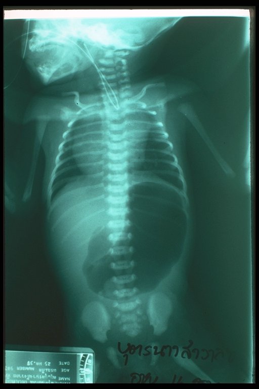
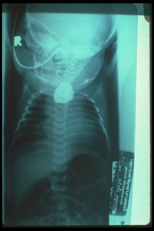
An X-ray finding in a baby with "triple atresia" : esophageal atresia, duodenal atresia and anorectal malformation. The nasogastric tube is coiled in the upper pouch. The typical distended stomach and upper duodenum indicated duodenal atresia is also noted |
|
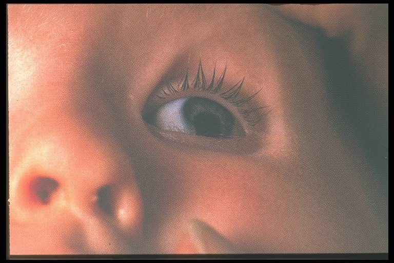
Coloboma, one of the multiple anomalies in CHARGE association. In such syndrome, esophageal atresia should be specifically sought. |
|
|
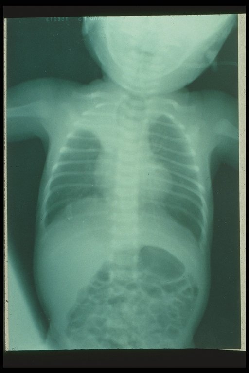
Chest X-ray showed atelectasis of the right upper lobe, this is the most common complication due to aspiration |
|
|
Chest X-ray revealed dilated blind proximal esophageal pouch and atelectasis of the right middle lobe |
|
|
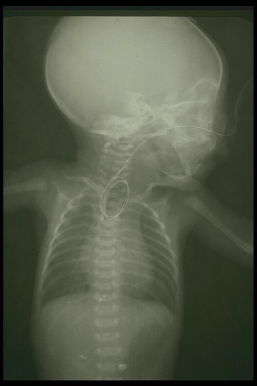
Anteroposteior film shows the coiled catheter in the blind upper pouch of esophagus. The air in stomach indicates that a distal tracheoesophageal fistula exists. |
|
|
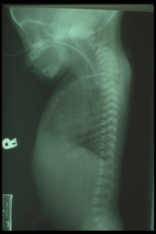
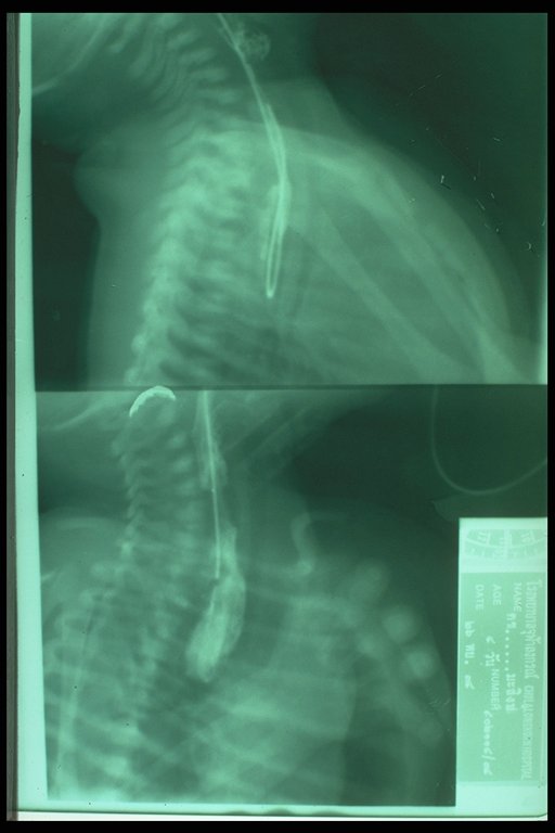
Lateral film shows the catheter in the blind upper pouch of esophagus. No air in the stomach indicates that there is no distal fistula |
|
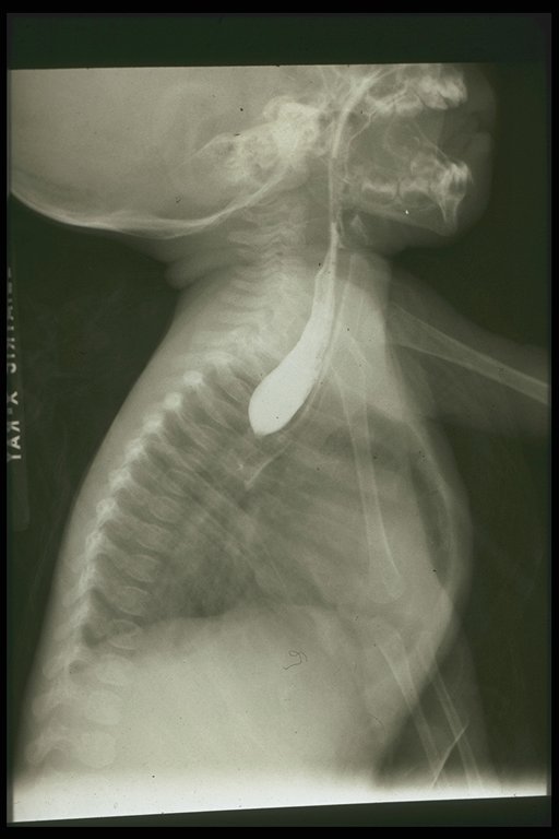
Plain lateral film reveals the coiled catheter in the esophagus. Contrast study proves that it is a blind pouch. |
|
|
Blind proximal esophageal pouch is demonstrated by contrast study (Lateral film) |
|
Blind proximal esophageal pouch is demonstrated by contrast study (Anteroposterior view) |

|
Blind proximal esophageal pouch is demonstrated by contrast study. Air in the stomach indicates the existence of distal fistula |