
|
|
| Course
Map: Tumor-like
Lesions: Radiation Cystitis |
| Radiation Cystitis | |
| Radiation therapy of any of the pelvic organs can result in tumor-like lesions of the bladder, months or years after such therapy [46, 51]. Microscopically, nodules of squamous epithelium push into the lamina propria without true invasive growth. The adjacent tissue is hemorrhagic and shows fibrin deposits and/or fibrinoid vascular changes. The deeper stroma often shows large, multinucleated stromal cells ("giant cell cystitis"). Four different cases are illustrated to show the repetitious histology of radiation cystitis. | |
| Previous Topic | Next Topic |
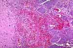 |
Figure 1: Rounded nests of squamous epithelium (right), hemorrhage (center), and large, reactive stromal cells (arrows) are typical features of radiation cystitis. |
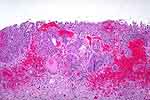 |
Figure 2: Aggregates of squamous epithelium and hemorrhage. |
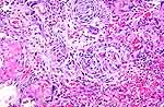 |
Figure 3: Radiation cystitis showing fibrin deposits (asterisks), hemorrhage (lower right), and squamous nests (center). |
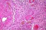 |
Figure 4: Fibrin deposits (asterisks) and squamous epithelium (higlighted), in addition to inflammation and vascular congestion. |