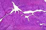
| Inflammatory lesions of the lamina propria, particularly when
accompanied by much edema, will produce an irregular mucosal contour with
broad-based or club-shaped surface projections (polypoid cystitis, Fig.
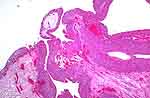
1). Sometimes, there will be a pronounced hyperplasia of the urothelium
with resulting exophytic excrescences resembling papillary neoplasms. This
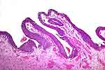
is shown in Figure 2 which is near a bladder fistula. More often the surface
protrusions will include both broad-based and narrow structures, so polypoid
and papillary cystitis are descriptive designations for a single entity.
Figures 3 and 4 show this variation. The delicate fibrovascular cores of
true neoplastic fronds are not present. [119].
|