
|
|
| Course
Map: Tumor-like
Lesions: Cysts |
| Cysts | |
| Clinically evident cysts will usually prove to be urachal cysts (Fig. 1), obviously located in or superior to the dome of the bladder. These may be lined by urothelium but colonic metaplasia is often present (Fig. 2, Fig. 3). These may be multilocular and may be associated with abscess or carcinoma. Rare cloacal cysts occur in the posterior bladder wall, and mullerian cysts occur between the bladder and rectum. Trigonal cysts are also rare. | |
| Previous Topic | Next Topic |
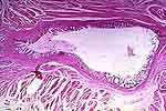 |
Figure 1: A small urachal cyst in the bladder wall. The lining epithelium shows glandular metaplasia. |
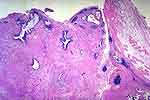 |
Figure 2: A large urachal cyst (arrows) with destroyed epithelial lining. Most of this view shows urachal remnants, some lined by urothelium, others by glandular metaplasia. |
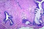 |
Figure 3: Same case as Fig. 2 showing urachal remnants. Such remnants should help to identify the nature of the cyst. One is lined by urothelium (arrow) and several by colonic epithelium, i.e. glandular metaplasia (arrowheads). |