
|
|
| Course
Map: Epithelial Tumors: Malignant: Non-urothelial
Carcinoma: Squamous Cell Carcinoma |
| Squamous Cell Carcinoma | |
| As we already noted, many transitional cell carcinomas will show focal or extensive areas of squamous metaplasia but the diagnosis of squamous cell carcinoma is made only when there is no urothelial element present. Most of these are well differentiated or moderately differentiated tumors meaning that there will be numerous keratin pearls or squamous eddies or intercellular bridges. Most of them will be found in the muscularis propria (pT2) at the time of initial diagnosis. The bladder epithelium near the tumor will generally show squamous metaplasia and it usually will be the keratinizing type of squamous metaplasia. This is, of course, the common type of carcinoma associated with schistosomiasis. In the United States most cases occur for no apparent reason but about 20\% are seen following some form of chronic irritation such as stones, chronic infection, chronic catheters or in diverticula. | |
| Previous Topic | Next Topic |
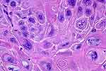 |
Figure 1: This is an invasive squamous cell carcinoma. Compared to urothelial carcinoma, the cells have an abundance of eosinophilic cytoplasm and clearly outlined cell borders. This photomicrograph was selected to illustrate, for the non-pathologists, the morphology of well differentiated squamous cells with unusually prominent intercellular bridges. |
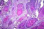 |
Figure 2: This is a well-differentiated squamous cell carcinoma. In the center, there is a large keratin pearl (arrow). |
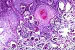 |
Figure 3: This is also a well differentiated squamous cell carcinoma with keratin pearls, and this example is associated with schistosomiasis. |
 |
Figure 4: Moderately differentiated squamous cell carcinoma. This field could be confused with a transitional cell carcinoma, but there is some keratin (arrow) in the center and the cell membranes are quite discrete. |
 |
Figure 5: Squamous cell carcinoma, pT2. Small, brightly eosinophilic keratin deposits are present (arrows), especially on the right but much of it is nonkeratinizing. This one would be diagnosed as moderately to well-differentiated squamous cell carcinoma. |