 |
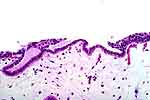
Figure 1: Cuboidal metaplasia with normal
urothelium on the right (arrow). |
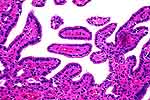 |
Figure 2: Papillary contour of the bladder mucosa
with all surfaces covered by a single row of cuboidal cells. |
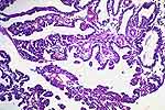 |
Figure 3: Papillary and polypoid contour of the
bladder mucosa with all surfaces covered by a single row of cuboidal
cells. |
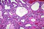 |
Figure 4: Tubules within the stroma have a
resemblance to renal tubules but this is a coincidental similarity - they
are not of nephrogenic origin. Larger tubules or microcysts are lined by
larger, hob-nail type epithelium. Lymphocytes and plasma cells occupy the
stroma. |
 |
Figure 5: Tubules across the center of the field
have prominent basement membranes. Lower left: plasma cells. |
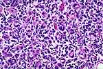 |
Figure 6: Extremely small tubules have prominent
basement membranes. Plasma cells are, again, a prominent feature. |
 |
Figure 7: The cytokeratin immunostain will
highlight and accentuate the morphology of the nephrogenic adenoma. |
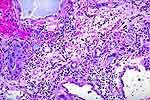 |
Figure 8: When the surgical procedure which
induced the formation of the NA was for carcinoma, one may see residual
carcinoma and NA in the same specimen. The carcinoma is on the left
(circle). |