HASHIMOTO'S THYROIDITIS
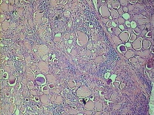
This thyroid shows scattered foci of dense lymphocyte infiltration. Note that thyroid follicles are in normal size (Fig.1).
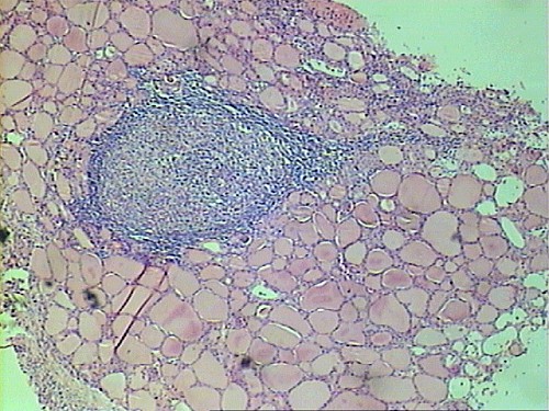
Germinal center (secodary lymphoid follicles) may present in some of the lymphoid infiltration (Fig.2).
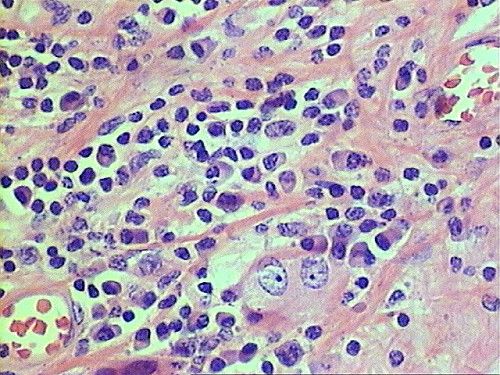
The lymphoid infiltration causes destruction and cellular change to the follicular cells within the area. The altered follicular cells may show enlarged nuclei and increase of eosinophilic granular cytoplasm (Fig.3).
This change is called oncocytic or Hurthle cell change. Compare the oncocytic follicular cells to normal follicular cells (Fig.4)( This change alone is not pathognomonic and can occur in various conditions).
Cellular infiltrate is composed of lymphocytes, and plasma cells in majority.
Note the two oncocytic cells that remain within the infiltrate (Fig.5).