Cysticercosis
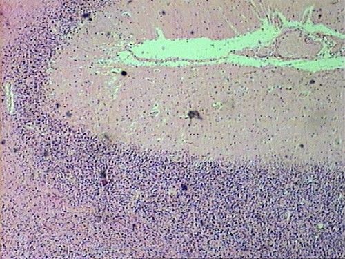
Sections show cerebellar tissue ( Fig.1).
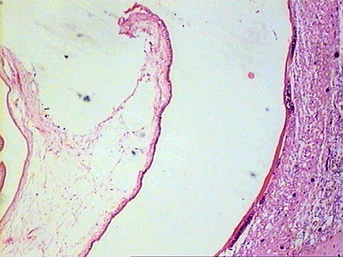
A cystic structure lined by a thin layer of fibrous tissue is seen ( Fig.2).
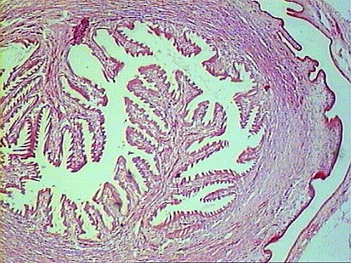
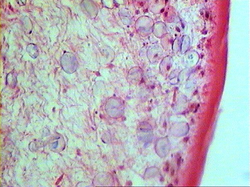
Within the cyst, the parasite is observed ( Fig.3, Fig.4).
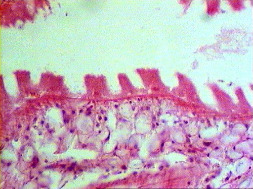
This thick cuticle with projection and calcareous bodies in the stroma are quite charateristic ( Fig.5, Fig.6).