Ẻ·´Êͺ Áըӹǹ 10 ¢éÍ ãËéãªéàÁéÒÊì¤ÅÔê¡Ë¹éҤӵͺ·Õè¶Ù¡µéͧà¾Õ§ 1 ¤ÃÑé§ â»Ãá¡ÃÁ¨ÐáÊ´§¤ÓµÍºÇèÒ ¶Ù¡ ËÃ×Í ¼Ô´ ¾ÃéÍÁ·Ñ駤Դ¤Ðá¹¹ãËé ËÒ¡µéͧ¡ÒäÓ͸ԺÒ¤ӵͺãËé¤ÅÔê¡·Õè

1.

This is a simple epithelium.
 
A. Can be either squamous, columnar or transitional?
B. Can be modified to cover the external body surface?
C. Can be keratinized?
D. Can be formed as the endothelium which lies lumens of heart and body cavities?
E. Can be formed as the mesothelium which lies lumens body cavities?
   
2. In a pseudostratified epithelium, …………………………………………….
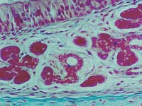
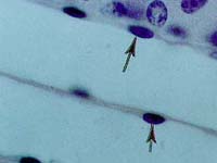
 
A. all cells rest on a basement membrane. 
B. all cells reach the free surface, but only some rest on the basement membrane.
C. all apical cell membranes are modified as microvilli.
D. there are two layers of cells, and only the basal cells rest on the basement membrane.
E. there are two layers of cells and two levels of nuclei.
   
3. If a membrane-impermeant fluorescent dye (MW <1,000) was injected into a single epithelial cell in the epidermis of the skin, you would expect it to appear in an adjacent epithelial cell due to …………………………………...
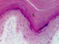
 
A. the presence of intercellar bridges between adjacent cells
B. the presence of tight-junctions between adjacent cells
C. diffusion across the plasma membrane
D. the presence of zonula adherens between adjacent cells
E. the presence of gap junctions between adjacent cells
   
4. Which epithelium (as seen in the photograph) can be distended to an extreme degree without losing its barrier function?
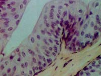
 
A. Stratified  squamous  non-keratinized  epithelium
B. Pseudostratified  epithelium
C. Transitional  epithelium
D. Simple  squamous  epithelium
E. All  of  the  above
   
5. Classify the epithelium that a physician first sees when examining a patient.
 
A. Simple squamous epithelium
B. Non-keratinized stratified squamous epithelium
C. Transitional epithelium
D. Keratinized stratified squamous epithelium
E. Pseudostratified columnar epithelium
   
6. Goblet cells are classified as ….………………  .
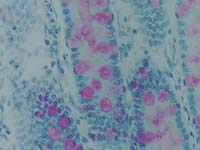
 
A. basal precursor cells
B. absorptive cells
C. holocrine-secreting cells
D. unicellular glands
E. paracrine gland
   
7. Mixed exo-endocrine gland as seen in the photograph is
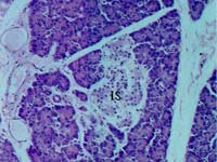
 
A. Islet of Langerhans
B. Seminiferous  tubules
C. Pancreas
D. Mammary gland
E. Crypts of Lieberkuhn
   
8. The serous demilunes are mostly found in the parenchyma of ……………………….  .
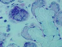
 
A. parotid  salivary  gland
B. submandibular  salivary  gland
C. Subligual  salivary  gland
D. apocrine  sweat  gland
E. exocrine  pancreas
   
9. The type of epithelium (as shown) is found in lining of ...........
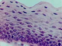
 
A. ureter
B. striated salivary duct
C. seminiferous tubule
D. vagina
E. renal tubule
   
10. The type of epithelium as seen in the screen, is ...................... epithelium ?
 
A. Pseudostratied ciliated columnar
B. Stratified ciliated columnar
C. Pseudostratied columnar, with stereocilia
D. Simple columnar, with microvilli
E. Simple columnar, with cilia
   
11. The  basement  membrane (BM) …………………………………  .
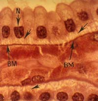
 
A. consists of a few connective tissue cells and reticular fibers.
B. consists mainly of collagen and elastic fibers
C. stains with PAS, indicating that it forms a continuous layer at the apical surface of an epithelium
D. cannot be seen in the light microscope by conventional staining
E. consists of basal lamina, protein-polysaccharides and reticular fibers
   
12. All of the followings are true for the apocrine sweat glands, except
 
A. They are found in the axilla.
B. The secretory units show apical caps or cytoplasmic buddings.
C. They are found in the palm and sole area.
D. Myoepithelial cells are present in the secretory units.
E. There are no centro-acinar cells at the secretory units.