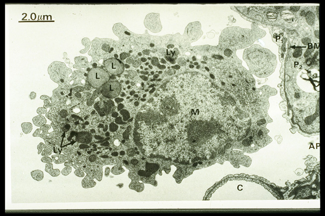
 Figure 126 :ภาพแสดง Alveolar macrophage (M) ระดับกล้องจุลทรรศน์อิเลคตรอน ซึ่งสัมผัสกับผนัง capillary (C) และ Type II pneumocyte (P2), AP= Alveolar pore และ BM = Basement Membrane กั้นอยู่ระหว่าง Type I (P1)& Type II pneumocyte(P2) ภายใน cytoplasm ของ alveolar macrophage บรรจุ secondary lysosomes (Ly) จำนวนมากและ lipid droplets (L)