| Department of Pathology, State University of New York at Stony Brook |

 Go to: TOC |
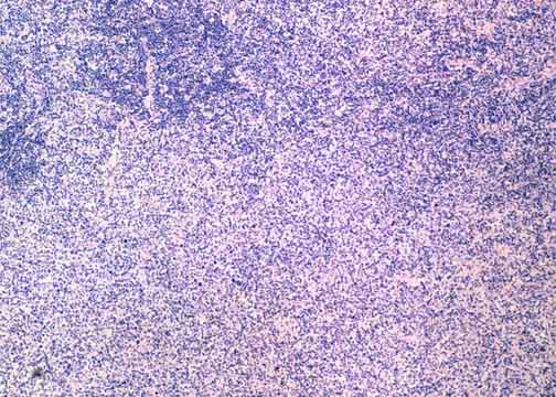
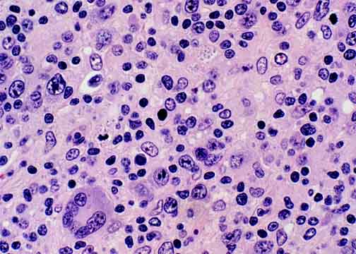
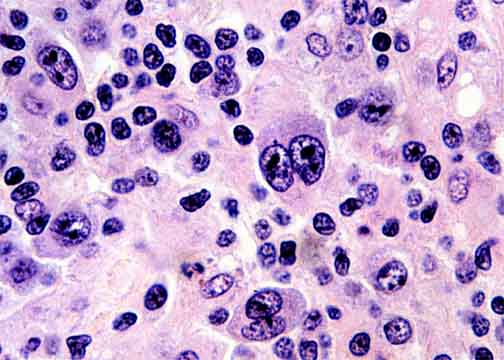
This lymphoma was also present in the bone marrow. Here in a lymph node it has a mixture of small and large cells. Some of the large cells show marked atypia including multinuclearity. Many scattered epithelioid histiocytes accompany the malignant T-cells, so that this case could be described as Lennert's lymphoma.
 Table of Contents |