Go to:

TOC
|
Peripheral T-Cell Lymphomas: Example 1
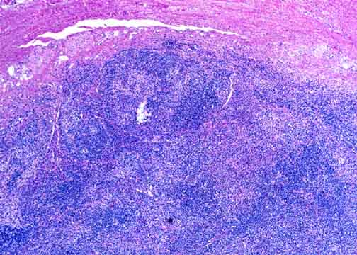
This is a large cell PTCL that partially replaced the lymph node. The case is unusual for the banded or mottled pattern seen here at low power. A thickened capsule is at the top. |
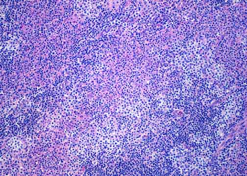
At medium power the bands are evidently composed of stripes of small lymphocytes (which may or may not be part of the lymphoma), stripes of large cells with clear cytoplasm, and stripes of pink-tinged necrosis with neutrophils. While this pattern is unusual and visually arresting, it does not have any significance beyond that of its constituent cells. |
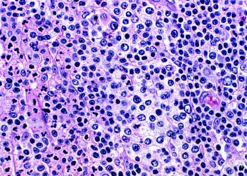
Here the neoplastic large T-cells with clear cytoplasm are seen in the center. The small, bluish lymphocytes are at the right, and the reddish areas of necrosis and neutrophils are at the left. |

Table of Contents 
|