ตัวอย่างที่ 3
![]()
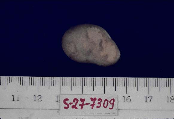
ภาพ Gross :
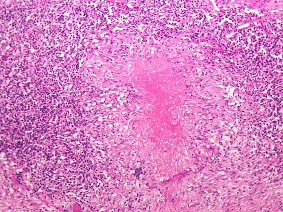
ภาพจากกล้องจุลทรรศน์ (microscopic feature)
 |
 |
| Formative Lab รายวิชา 3000369 Foundation of Pathology Diseases ปีการศึกษา 2558 | ||
| ชื่อ______________________นามสกุล______________________เลขประจำตัว____________________เลขที่________ | ||
| อวัยวะ (organ) (1 คะแนน) ..................Lymph node..................................... | ||
| บรรยายพยาธิสภาพ gross ของรอยโรคจากภาพ (3 คะแนน) .....A lymph node measures 2.5X2 cm. in dimension. The cut surface is solid and grey-white with areas of yellowish map-like lesion. | ||
| ต่อมน้ำเหลืองมีขนาด 2.5X2 ซม. หน้าตัดทึบ มีสีขาวเทา ปะปนด้วยรอยโรคมีลักษณะเป็นแผ่นที่สีเหลือง | ||
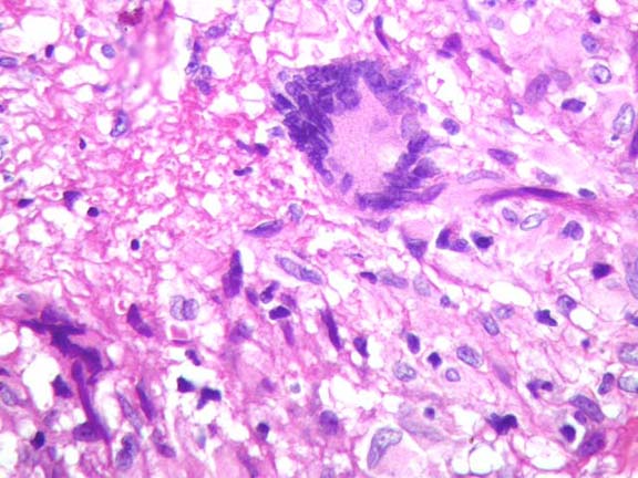
| บรรยายพยาธิสภาพ จากกล้องจุลทรรศน์ ของรอยโรค (3 คะแนน) A well-defined granulomatous lesion comprising aggregation of epitheloid cells (macrophage) and a central caseous necrosis. Scattering among the epitheloid cells are multinucleated giant cells (called Langhan giant cell) | ||
| พบการสะสมของ epitheloid cell ในรอยโรคที่มีลักษณะเป็นแผนที่สีเหลืองของต่อมน้ำเหลือง ทำให้เกิดเป็นก้อนที่มีขอบเขตที่ชัดเจน (เรียกว่่า granuloma) ตรงกลางก้อน มีการสะสมของกลุ่มเนื้อตาย ที่เรียกว่า caseous necrosis นอกจากนี้ยังพบเซลล์ขนาดใหญ่ที่มีหลายนิวเคลียส์ เรียกว่า multinucleated giant cell (หรือเรียกว่า Langhan's giant cell) ภายในก้อน granuloma นี้ | ||
| วินิจฉัยรอยโรค (Diagnosis) (1 คะแนน) | Granulomatous lymphadenitis. | |
| หรือ | Granulomatous inflammation with caseous necrosis | |
| เหตุผลในการวินิจฉัย (1 คะแนน) : Aggregation of epitheloid cells with caseous necrosis. | ||
| พบการสะสมของ กลุ่มเซลล์ epitheloid และตรงกลางก้อนพบ เนื้อตายชนิด caseous necrosis | ||
| อาการหรือการตรวจพบที่สำคัญ (Chief complaints) (1 คะแนน) : Palpable lymph node | ||
| คลำต่อมน้ำเหลืองได้ | ||
![]()