
|
|
| Course Map:
Miscellaneous Tumors: Melanoma |
| Melanoma | |
| Primary melanomas of the bladder are rare and do not differ histologically, biologically, or by immunohistiochemistry from those seen elsewhere [3,62,106]. Many patients dying of malignant melanoma have bladder metastasis and when considering a diagnosis of primary vesicle melanoma, certain criteria should be met: no history of prior cutaneous, opthalmic, or other melanoma should be evident; examination of all skin surfaces should be negative, and the distribution of any metastatic deposits should be consistent with a tumor originating in the bladder. Atypical melanocytes in the bladder epithelium will obviously support a diagnosis of primary melanoma. | |
| Previous Topic | Next Topic |
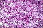 |
Figure 1: Poorly differentiated tumor cells are arranged into rounded aggregates. The diagnosis is revealed mostly by the melanin pigment seen in Figure 2. This might initially suggest a nested urothelial carcinoma, but note that some of the cells have a ballooned appearance or clearing of the cells peripherally. |
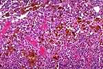 |
Figure 2: Same case as figure 1. Melanin pigment is present. |
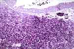 |
Figure 3: This is another case, demonstrating the positive HMB-45, diagnostic of melanoma. |
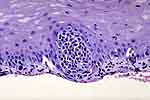 |
Figure 4: In this case the melanoma (not shown) was in the urethra. This is the adjacent surface epithelium, and it shows atypical melanocytes (arrows) arranged individually (lower left) and in rounded nests. The latter vary markedly in size (circles) (Melanoma in-situ). |