
|
|
| Course
Map: Epithelial Tumors: Malignant: Urothelial Carcinoma:
Carcinoma In-situ |
| Carcinoma In-situ | |
| This is regarded as a precursor to invasive carcinoma, at least in many cases [8]. It is defined as a "flat" or nonpapillary surface epithelium that contains any number of cytologically malignant cells. This represents a significant change from a previously held view that in-situ carcinoma required that the full thickness of the epithelium contain cytologically malignant cells. Nests of von Brunn may also show CIS, with or without involvement of the surface urothelium. Malignant cells sometimes are seen only in the lower part of the epithelium or sometimes in only the surface layer. In the pagetoid type of CIS, small groups or individual malignant cells are scattered through an otherwise normal epithelium. [80]. The "clinging" type of CIS results from desquamation of most malignant cells, spontaneously or from trauma, leaving but few anaplastic cells attached to the basement membrane. Some observers may recognize this present definition of CIS as including those lesions previously diagnosed as "severe dysplasia" and also some cases of "moderate dysplasia". The critical element here is the recognization of cytologic malignancy. One will occasionally see the term "high grade intraurothelial neoplasia" used instead of CIS. | |
| Previous Topic | Next Topic |
 |
Figure 1: Carcinoma in-situ. The nuclei are dark-staining and they vary markedly in their size and shape and involve the full thickness of the epithelium. |
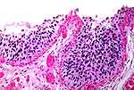 |
Figure 2: Carcinoma in-situ involving surface(left) and a von Brunn nest (right). This was photographed at the same magnification as Figure 1, and one can see that cellular anaplasia is less extreme, but the same comment about nuclear morphology is applicable. |
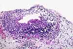 |
Figure 3: Carcinoma in-situ involving a von Brunn nest. The anaplastic nuclear characteristics described in Figure 1 and Figure 2 involve only a part of this nest. |
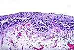 |
Figure 4: Carcinoma in-situ. Only the lower half of the epithelium is involved. |
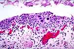 |
Figure 5: The pagetoid type of carcinoma in-situ. Malignant cells (highlighted) appear to be invading through an epithelium that is otherwise normal. |
 |
Figure 6: A few cells with malignant nuclear features (large, dark, varied size and scant cytoplasm) remain attached to the basement membrane (arrows): the "clinging" type of carcinoma in-situ. |